Q4E Case Study 20
- Anatomical Movements
Proposed Subject usage:
Sport and PE (GCSE/AS / A level)
| |
National
Curriculum (Key Stage 4) |
|
| |
3. Evaluating
and improving performance. |
|
| AQA
GCSE PE Specification |
6.2 Analysis of performance
The specification will assess a candidate’s ability to
analyse performance so as to:
- Determine its strengths and weaknesses
- Improve its quality and effectiveness
|
| AQA
A Level PE Specification |
At AS, candidates
are required to observe, analyse and evaluate performance.
21.2 Observe the chosen performer in relation to the competent
performance of the 5 specific techniques for a chosen activity |
Introduction
In order to perform a practical analysis of human movement a sound understanding of anatomical movements is necessary. Anatomical movements can be defined as the act or instance of moving the bodily structures or as the change of position in one or more of the joints of the body. Joint actions are described in relation to the anatomical position which is the universal starting position for describing movement. A subject is considered to be in the anatomical position when they are standing in an upright posture, facing straight ahead, with their feet close together and parallel and the palms of their hands facing straight ahead. This position is demonstrated in Figure 1 below.
When studying the various joints of the body and analyzing their movements it is helpful to characterize them according to specific planes of motion and their axes. A plane of motion may be defined as an imaginary two-dimensional surface through which a limb or body segment is moved. In the human body there are three planes of motion (Figure 1) in which the various joint movements can be classified. Similar to the planes of motion the axes of rotation may be considered as a series of imaginary lines that run through the body; there are also three axes of rotation (Figure 2) where movement can occur.
- Sagittal (anteroposterior) plane – This plane is vertical and bisects the body from front to back. Dividing it into right and left symmetrical halves. For movement to occur in the sagittal plane rotation about the horizontal axis (transverse axis) must take place.
- Frontal (coronal) plane – This plane bisects the body laterally from side to side, dividing the body into front and back halves. Movement in the frontal plane takes place about the anteroposterior axis (frontal axis) must take place.
- Transverse (horizontal) plane – This plane divides the body horizontally into superior and inferior halves. Movement in this plane takes place about the longitudinal axis (vertical axis).

Objectives
- To define a number of anatomical movements and demonstrate these movements with appropriate illustrations using the Quintic software.
- Identify sport specific skills where the identified anatomical movements occur and determine the role these movements play in successful completion of the sport skill.
Methods
- Video footage was captured at 50 fps using a Panasonic 3CCD Camera of a subject performing anatomical movements. The videos were then exported into Quintic Biomechanics 9.03v17 software.
- The blend, shapes and still capture functions in Quintic were used to illustrate the various anatomical movements from the captured footage.
- Video footage was then captured of various sport skills and opened in the Quintic software where they were analysed in order to determine the specific joint movements that the skill was composed of.
Functions of the Quintic software used:
- Single Camera Function
- Still Image Capture
- Photo Sequence module
- Shape Tools
- Blend Function
- Play Speed Function
Results
Note: All movements being described assume the body begins from the anatomical position unless stated otherwise as described in the introduction.
1. Flexion
Flexion is a bending movement that results in the decrease of the angle in a joint by bringing bones closer together. It usually occurs in the sagittal plane. The table below represents some of the joints where flexion can occur and an example of that motion;
Flexion |
Joint |
Example |
Shoulder |
Raising your arms upwards and in front of the body. |
Elbow |
Movement of the forearm to the shoulder by bending the elbow to decrease its angle |
Spine |
Moving the chin towards the chest. |
Hip |
Movement of the femur towards the pelvis / bringing knee into chest |
Knee |
Moving the heel towards the buttocks. |
Figure 3 below demonstrates flexion of the wrist joint. Flexion of the wrist joint is an important movement in many sport skills especially racquet sports as it can provide stability and power to a performance. In the follow through phase of a basketball free-throw wrist flexion is evident. The purpose of this movement which is technically known as a wrist snap (demonstrated in Figure 4 below) is to generate enhanced spin on the ball which will in turn add lift to the trajectory (flight of the ball), this can increase release velocity of the ball and improve the overall performance making it an important aspect to the execution of a basketball free-throw.

2. Extension
This is a straightening movement that results in the increase of the angle in a joint by moving bones further apart. This movement occurs in the sagittal plane and is the reverse movement to flexion. The table below represents joints of the body that extension can occur in and an example;
Extension |
Joint |
Example |
Shoulder |
From flexion lowering your arms downwards and in front of the body. |
Elbow |
Following flexion it involves movement of the forearm away from the shoulder by straightening the elbow. |
Spine |
Moving the head backwards so that you would finish looking straight up. |
Hip |
From flexion returning the femur to anatomical position |
Wrist |
Movement of the hand towards the back of the forearm |
Figure 5 demonstrates the knee joint performing extension. The knee joint is the largest joint in the body and is primarily concerned with weight bearing and locomotion, for these reasons knee extension is a part of numerous sport skills. Any sport that requires jumping relies greatly on knee extension for successful completion of the jump. Running is one of the most basic skills where knee extension can be seen (Figure 6 shows the knee in extension). During the drive phase of running the drive leg extends at the knee joint in order to generate forward and upward thrust which propels the body making knee extension a principle component of running.

3. Abduction
This is a lateral movement away from the midline of the trunk and it occurs in the frontal plane. The table below demonstrates the joints in the body where abduction can occur along with an example;
Abduction |
Joint |
Example |
Shoulder |
Upward lateral movement of the humerus out to the side. |
Hip |
Movement of the femur in the frontal plane laterally to the side away from the midline |
Wrist |
Movement of the thumb side of the hand toward the lateral aspect of the forearm |
Abduction at the hip joint is shown in Figure 7 below. Some cricket bowlers demonstrate hip abduction during a fast bowl as illustrated in Figure 8 below. In order to produce high ball release speeds, fast bowlers require high joint torques. High joint torques can be created through counter rotation which is rotation of the upper trunk away from the direction that the ball is to be thrown. By abducting the hip joint as well as other movements counter action can be created which will increase the joint torques and subsequently increase the release speed of the ball. Also from the position shown in Figure 7 below the hip abduction creates a greater distance for trunk rotation to occur over which allows more momentum and power to be created.

4. Adduction
This is a movement medially toward the midline of the trunk and it occurs in the frontal plane. The table below illustrates the joints where adduction can be performed with examples for each joint;
Adduction |
Joint |
Example |
Shoulder |
From abduction it is the downward movement of the humerus in the frontal plane medially toward the body. |
Hip |
From adduction it is the movement of the femur in the frontal plane medially toward the midline or putting your leg across your body. |
Wrist |
Movement of the little finger side of the hand toward the medial side of the forearm. |
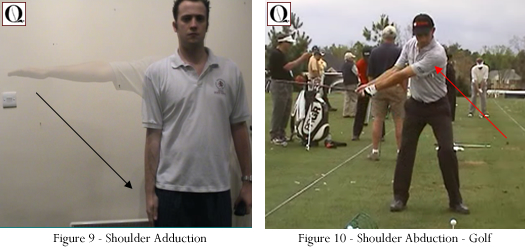
Figure 9 illustrates the shoulder joint performing adduction. Shoulder adduction is a key movement in the performance of a golf swing and is illustrated in Figure 10 below. During a golf swing the body segments work together in a coordinated sequence to maximize club head speed at ball impact. The lead arm accelerates into maximum adduction during the backswing which allows the golfer to attain a suitable position for the upcoming downswing and hence increases efficiency of the overall skill.

5. External Rotation
This is a rotary movement around the longitudinal axis of a bone away from the midline of the body. This movement occurs in the transverse plane and is also known as rotation laterally, outward rotation and lateral rotation. The joints in which external rotation can occur are shown in the table below;
External Rotation |
Joint |
Example |
Shoulder |
Movement of the humerus laterally in the transverse plane along its long axis away from the midline |
Hip |
Lateral rotary movement of the femur in the transverse plane around its longitudinal axis and away from the midline. |
Knee |
Rotary movement of the lower leg laterally away from the midline. |
Figure 11 below demonstrates external rotation of the shoulder joint. A sporting example where external rotation is evident is during the tennis serve, this is illustrated in Figure 12 below. During the wind up of the tennis serve the shoulder joint performs external rotation in order to store elastic potential energy which can be transferred into the forward motion of the serve which will generate a tremendous amount of force and momentum. Also external rotation allows the racquet to go further down, this is important because it will firstly allow more strain energy to be formed and secondly increase the acceleration time of the racquet during the forward phase allowing increased time for the transfer of energy.

6. Internal Rotation
This is a rotary movement around the longitudinal axis of a bone toward the midline of the body. It occurs in the transverse plane and is also known as rotation medially and inward rotation. The joints of the body where internal rotation can occur are listed in the table below as well as an example for each joint;
Internal Rotation |
Joint |
Example |
Shoulder |
Movement of the humerus in the transverse plane medially along its long axis toward the midline |
Hip |
Medial rotary movement of the femur in the transverse plane around its longitudinal axis and towards the midline. |
Knee |
Rotary movement of the lower leg medially toward the midline. |
Internal rotation is a very common movement in the human body. When we walk or run our hips undergo internal rotation with every stride we take. The run up in javelin demonstrates internal rotation as illustrated in Figures 13 and 14 below. The purpose of this movement is the production of momentum which can be transferred to the hand and wrist in order to maximize release speed of the javelin. Furthermore the consequence of the internal rotation of the hip is forceful rotation of the trunk which produces momentum which can be transferred to the upper body and used. For these reasons internal rotation of the hip joint is an important factor in the successful completion of a javelin throw.

Conclusion
A sound knowledge of anatomical movements is necessary in order to perform a practical analysis of human movement. Each anatomical movement occurs in a specific plane and around a specific axis of rotation. By examining sport skills you can determine the individual anatomical movements of which the skill is composed of and furthermore evaluate the influence the individual movement has on the overall success of the skill. This allows a greater knowledge of the skill to be achieved and the components that make that skill a success.
Downloads
Written
Case Study
|

~3.35 MB |
|

